
To the performance overview
PET-CT in prostate cancer (PSMA-PET/CT)
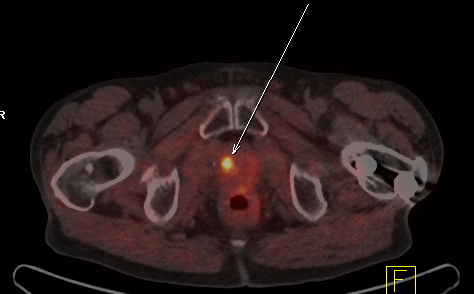
Figure: Local recurrence of a prostate carcinoma after removal of the prostate
PET/CT is currently the most advanced imaging method for tumour detection. It combines the advantages of positron emission tomography (PET) and computed tomography (CT) in a single device. The PET can display the tumour metabolism, whereas the CT can localize where exactly the tumour is. This allows to detect even the smallest tumours at a very early stage, which has not been possible with other methods.
In the PET study, patients receive a small amount of a low-radiation marker substance, PSMA or choline. This substance will concentrate in the cells of the prostate carcinoma.
On the PET image the diseased cells can be recognized as luminous dots. At the same time, the interior of the body is shown three-dimensionally by using computer tomography.
In the case of prostate cancer, the PET/CT examination might be necessary if the re-emergence of the cancer is suspected, if the tumour marker PSA increases, if prostate cancer is suspected although the biopsy is negative or if metastases in lymph nodes or other organs are suspected.
The early diagnosis allows the treating physician to choose the best treatment for the patient. A PET/CT follow-up can show whether the treatment is effective and the patient responds to the treatment e.g. chemotherapy.
Prostate carcinoma is the most common tumour disease in men in Europe. In Germany, tens of thousands of men are affected every year. Early diagnosis increases the chance of recovery.
Examination procedure
First a small amount of a low-radiation marker substance will be injected. This substance will accumulate in metabolically active tissue.
The substances used are very well tolerated; allergic reactions are not known. The radiation load is low and quickly degraded.
PET/CT images from the pelvis are then recorded for approximately ten minutes.
After the injection of the substance, you relax for about an hour in a relaxation room.
Then pictures of the whole body will be taken, which takes about 15-30 minutes.
During recording you will lie on a narrow examination table, that slides into and out of the round whole of the PET/CT unit. The opening is quite large and the tunnel is so short that you do not have to feel constricted. The measurement proceeds almost noiselessly.
The examination takes a total of about two to three hours.
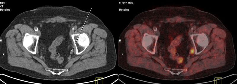
Pelvic lymph node metastases in the pelvis in a patient with prostate carcinoma
Preparation before and after the examination
You are allowed to eat on the day of the exam.
Please drink at least half a litre of water before testing.
You can take your medication as usual, however we would kindly ask you to bring your medication list with you.
Please also bring the results of your preliminary examinations. Particularly important are print-outs or the CD of the most recent CT and MRI examinations.
The radiotracer is individually ordered for each patient and is not storable (the costs run several hundred euros). Hence we kindly ask you to always keep your appointment on time and if for any reason you need to reschedule or cancel your appointment please do this at least two days in advance.
The pictures and the findings are evaluated within one day and sent to your treating physician as soon as possible. You will get a CD with the PET/CT images.
Please do not hesitate to contact us if you have any further questions!
Download “Patient Information PET-CT in Prostate Cancer” (PDF)