To the performance overview
Bone scintigraphy
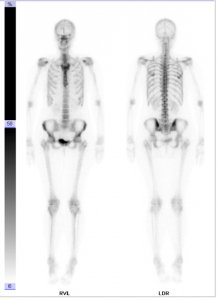
The skeletal or bone scintigraphy is a nuclear medical examination which can be used to detect pathological changes in the skeleton. For this purpose, a small amount of a low-radiation marker substance is administered. The substance accumulates in the bone structure and emits radiation. The radiation can be recorded as picture (scintigram) with a gamma camera.
It is possible to detect abnormal changes of the skeleton such as inflammation, bone fractures, signs of wear, bone tumours or bone metastases of a tumour at a very early stage due to this examination. It also detects loosening of joint prostheses.
Examination procedure
First, a small amount of a low-radiation marker substance will be injected. This substance will accumulate in areas where the metabolic level of the bones is elevated.
The substances used are very well tolerated; allergic reactions are not known. The radiation load is low and quickly degraded.
After that, first images of the skeleton are taken with a gamma camera for about twenty minutes.
Images of the skeleton will be taken once again about two to three hours later, as the metabolism of the bones is relatively slow.
The examination lasts approximately three to four hours, including waiting time. You do not have to stay at the practice while waiting. You may go home, to work or go at the cafeteria.
Preparation before and after the examination
You may have meals in advance to the examination, as well as take your medication as usual.
After the injection of the low-radiation marker substance, please drink at least half a litre of water.
Please empty your bladder shorty before the pictures are taken. Patients with urine bags should replace the bag with a new one.
We will send the examination results to your treating physician as soon as possible. He will then contact you to discuss the results as well as the possibility of a treatment.
Please do not hesitate to contact us if you have any further questions!


